Laboratory Microscopes for Cell Imaging and Research
Laboratory microscopes are optical instruments that form magnified images of specimens too small to resolve with the naked eye, enabling visualization of cells, tissues, microorganisms, and structural details at magnifications from 7x (stereo) to 1000x and beyond (compound or fluorescence). Types for research laboratories include inverted microscopes for cell culture observation in dishes and microplates, upright compound microscopes for fixed-tissue and histological slides, stereo microscopes for dissection and surface inspection, and fluorescence microscopes for imaging labeled cells and proteins.
MBP provides purchase order procurement for laboratory microscopes for research institutions across the USA and Canada. Request a quote for laboratory microscopes for cell imaging, microscopy, and life science research by contacting customerservice@mbpinc.net.
Microscopes

Showing 1 to 12 of 22 results
What Are Laboratory Microscopes?
Laboratory microscopes use optical lenses and illumination systems to produce magnified, high-contrast images of specimens, enabling researchers to observe cellular and subcellular structures, tissue morphology, surface features, and fluorescently labeled molecules. The four most common types in life science research are: inverted microscopes (objectives below the stage, sample viewed from beneath, the standard for cell culture in dishes and flasks); upright compound microscopes (objectives above the stage, used for fixed slides and smear preparations); stereo microscopes (dual optical path, low-magnification three-dimensional view for dissection and manipulation); and fluorescence microscopes (any configuration with epi-fluorescence illumination and matched filter sets for imaging fluorescent dyes and reporter proteins). Contrast methods include brightfield (standard), phase contrast (for unstained living cells), differential interference contrast (DIC, for optical sectioning), and darkfield (for surface detail on opaque specimens). LED illumination systems have largely replaced traditional mercury and halogen sources across all microscope classes, providing longer lamp life (above 50,000 hours), lower heat output, and instant-on operation. For live mammalian cell culture, specify an inverted microscope with phase contrast and at a minimum one fluorescence channel. For histology and fixed slides, specify an upright compound microscope. For dissection and manipulation, specify a stereo microscope.
What will you find :
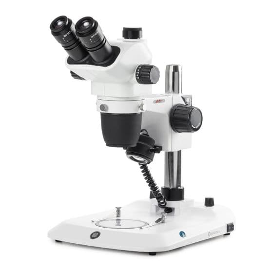
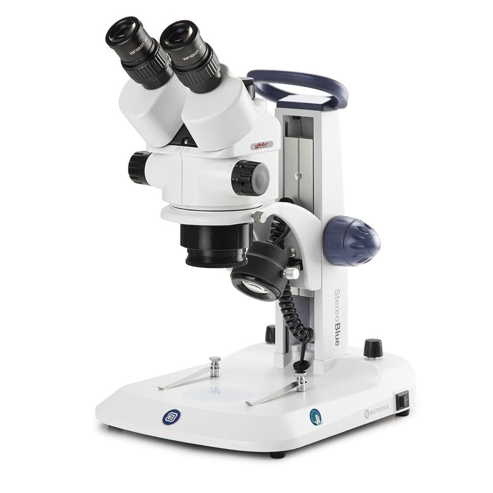
- Stereo Microscopes: Three-dimensional optical microscopes designed for low-magnification viewing, inspection, dissection, and manipulation of specimens using surgical tools and precision instruments.
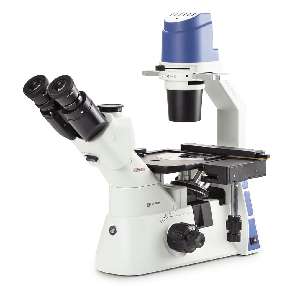
- Inverted Microscopes: Specialized microscopes with the objectives positioned below the stage, enabling observation of living cells and tissues in culture dishes, flasks, and microplates without disturbing the sample.
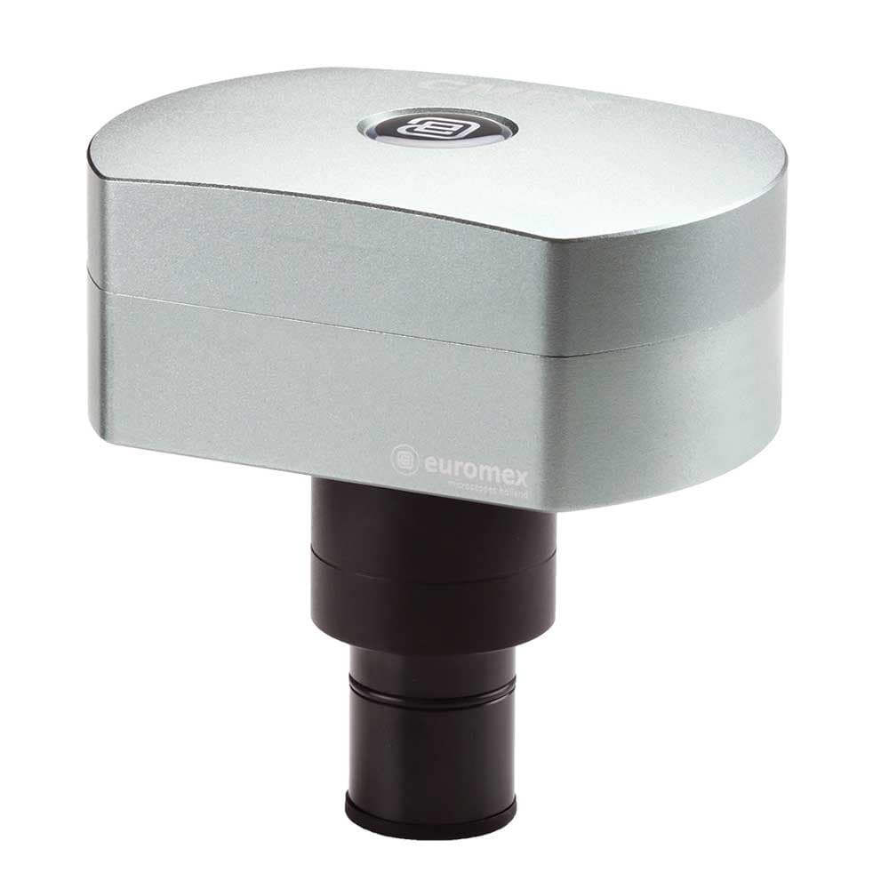
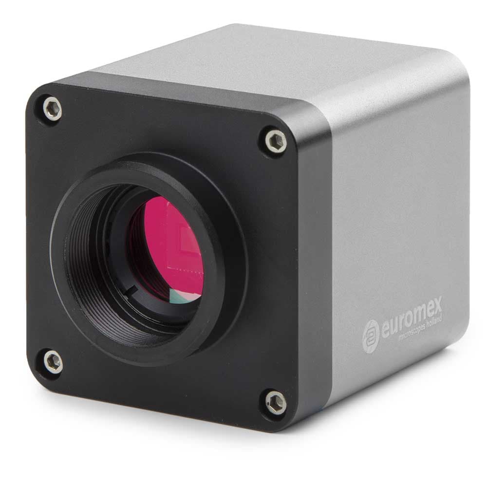
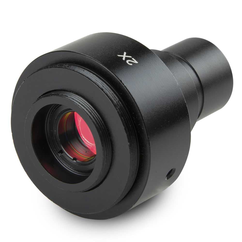
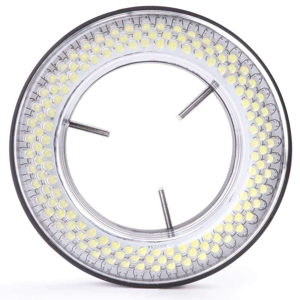
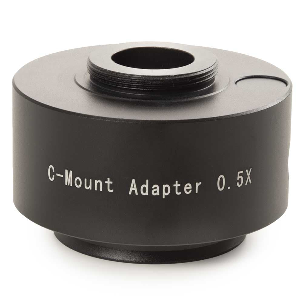
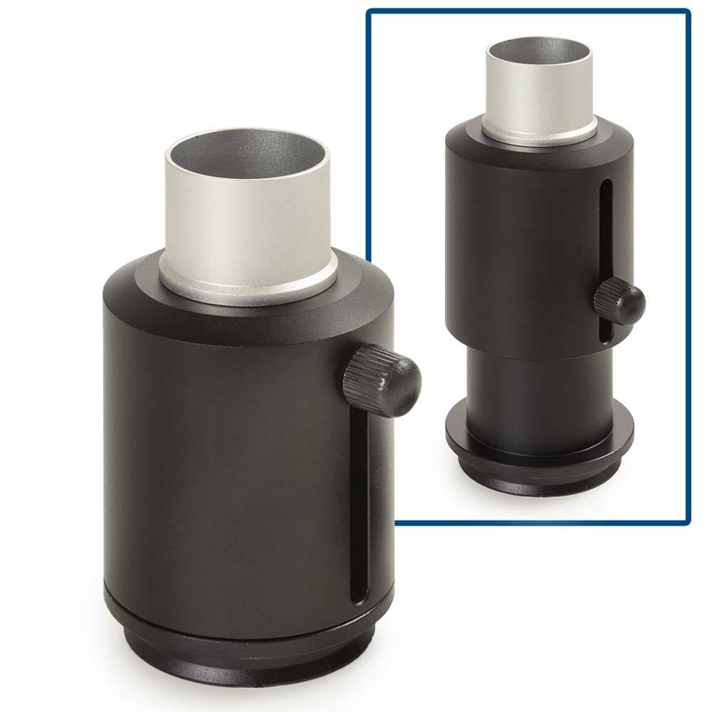
- Microscope Accessories: Supporting components that enhance microscope functionality, including digital imaging cameras, objective lenses, illumination systems, adapters, and other application-specific accessories.
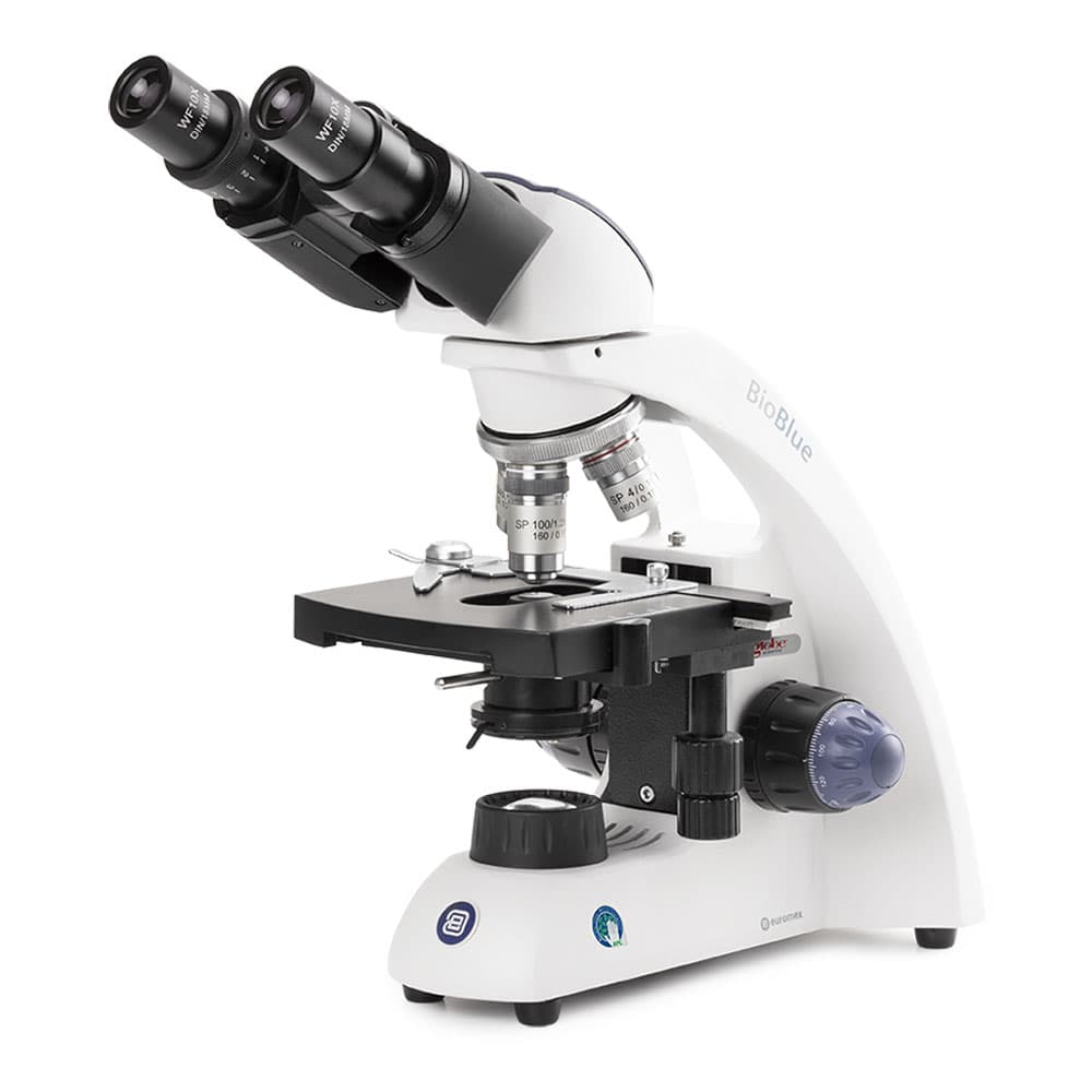
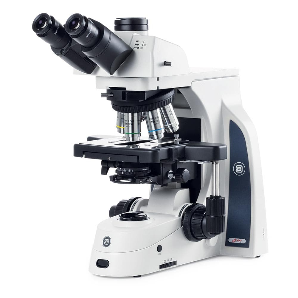
- Compound Microscopes: Multi-lens optical microscopes used for high-resolution examination of transparent or stained specimens such as cells, tissues, microorganisms, and histological samples.
How to Choose a Laboratory Microscope
Inverted vs. upright configuration
Inverted microscopes position the objective lens below the stage and the illumination source above, allowing observation of cells growing at the bottom of culture dishes, flasks, and microplates without disturbing the culture. They are the standard for mammalian cell biology, cancer research, developmental biology, and any application where cells are grown in standard tissue culture vessels. Upright microscopes position the objective above the stage and illuminate from below through the specimen, requiring thin, translucent samples mounted on glass slides; the standard for histopathology, microbiology smears, and cytology.
Objective lenses: magnification and numerical aperture
Objective lenses determine magnification, resolution, and working distance. Numerical aperture (NA) is the key specification for resolution: a higher NA provides finer resolving power. Standard objective sets for cell biology: 4x (NA 0.10) for overview; 10x (NA 0.25 to 0.30) for confluency assessment and colony counting; 20x (NA 0.40 to 0.45) for cell morphology detail; 40x (NA 0.60 to 0.75) for subcellular detail; 60x or 63x oil immersion (NA 1.25 to 1.40) for highest-resolution fluorescence imaging. Confirm the objective's working distance is compatible with the culture vessel bottom thickness; standard tissue culture plasticware has a 1 to 1.2 mm bottom thickness.
Phase contrast for cell culture
Phase contrast is the essential contrast method for observing unstained, transparent living cells in culture. Phase contrast objectives pair with a matched annular phase ring in the condenser (inverted) or below the stage (upright), producing contrast from differences in the refractive index of cell structures rather than from staining. Without phase contrast, mammalian cells in culture are nearly invisible against the background in brightfield. Confirm that the phase contrast objective designation (PhI, PhII, PhIII for low, medium, high contrast) matches the phase ring installed in the condenser.
Fluorescence: light source and filter sets
Fluorescence microscopes use an excitation light source (mercury lamp, LED, or laser) and matched filter cubes (excitation filter + dichroic mirror + emission filter) to selectively excite fluorescent dyes and reporter proteins and image their emission. LED fluorescence systems offer the longest source life (above 50,000 hours), fastest switching between channels, and lowest heat output, and are the standard for new purchases. Common filter sets: DAPI (UV excitation, blue emission), FITC/GFP (blue excitation, green emission), TRITC/mCherry (green excitation, red emission), Cy5/AlexaFluor647 (red excitation, far-red emission).
Camera and image capture
All research microscopes benefit from a camera port and a digital camera for image capture, documentation, and analysis. CMOS cameras with 2 to 20 megapixel resolution cover most brightfield and fluorescence applications. Scientific CMOS (sCMOS) and cooled CCD cameras provide lower noise for low-light fluorescence imaging of dim samples. Confirm camera mount compatibility (C-mount is standard) and required image analysis software before purchasing the camera system.
Specifications Context
Microscope optical quality is characterized by the optical system (finite vs. infinity corrected) and the objective correction class (plan-achromat for flat, color-corrected field across wavelengths; plan-apochromat for superior color and spherical correction used in fluorescence). Infinity-corrected optics are standard on all research-grade microscopes since approximately 2000 and allow accessories (filter cubes, cameras, motorized stage modules) to be inserted in the infinity space between objective and tube lens without compromising image quality. Finite-optics microscopes are older or entry-level systems with more limited expandability.
Contact the MBP team to request a quote and find the right microscope solution for your laboratory.
FAQ