Stereo Microscopes for Dissection and 3D Specimen Imaging
Stereo microscopes (also called dissecting or stereoscopic microscopes) use two independent optical paths to produce a three-dimensional, upright view of solid, opaque, or thick specimens. They are commonly available in magnifications ranging from approximately 7× to 45× on standard zoom models, with higher magnifications available on select configurations. Their long working distance provides ample clearance for specimen manipulation during dissection, electronics inspection, quality control, and other laboratory and industrial applications.
MBP provides purchase order procurement for stereo microscopes for research institutions across the USA and Canada. Request a quote for stereo microscopes for dissection, specimen inspection, and 3D imaging applications by contacting customerservice@mbpinc.net.
Stereo Microscopes
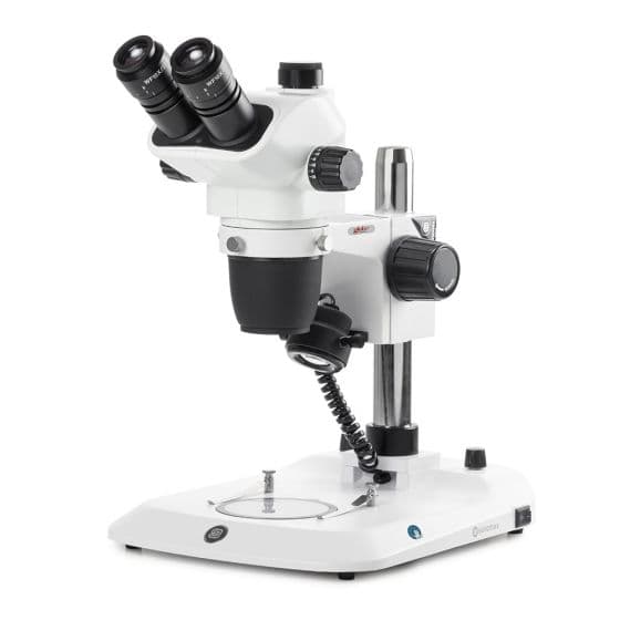
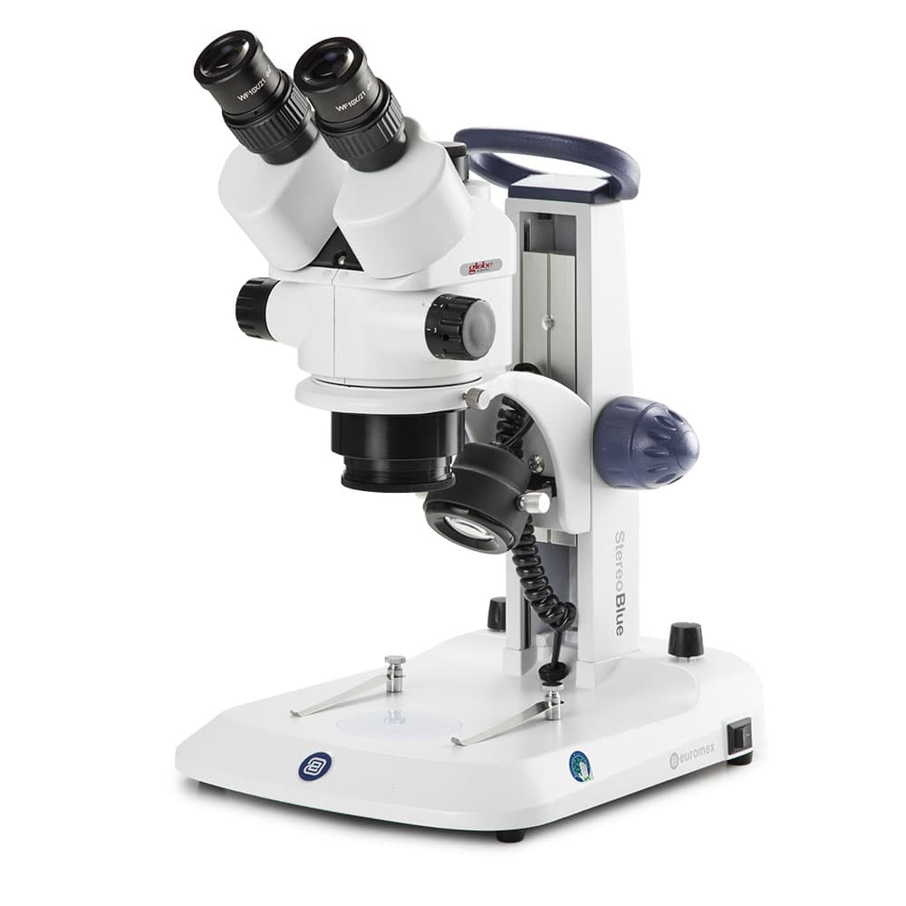
Showing 1 to 2 of 2 results
What Are Stereo Microscopes?
A stereo microscope uses two separate optical paths (each slightly offset from the other) to present a slightly different angle of the specimen to each eye, producing true stereoscopic (three-dimensional) depth perception in the image. Unlike compound microscopes, which use transmitted light through thin, translucent, slide-mounted specimens at high magnification, stereo microscopes use incident (reflected) illumination to observe solid, opaque, or thick specimens from above at lower magnifications. This design provides two key practical advantages: a long working distance (typically 80 to 115 mm at 1x objective) that allows tools, needles, forceps, or soldering irons to be operated under the lens during observation; and a wide field of view that allows large specimens (whole insects, embryos, plant organs, PCBs, rocks) to be seen in context. Stereo microscopes are used in biology (dissection, organism manipulation, embryo handling, zebrafish imaging), materials science and electronics (PCB inspection, solder joint analysis, microelectronics assembly), geology and paleontology (fossil and mineral examination), medical device assembly, and quality control inspection. Use a stereo microscope when you need to observe or manipulate a solid, opaque, or thick specimen in three dimensions with tools beneath the lens; it is the correct instrument for dissection, inspection, and manipulation, not a compound microscope.
How to Choose a Stereo Microscope
Zoom ratio and magnification range
Zoom ratio (the ratio of maximum to minimum magnification in the continuous zoom range) determines how much flexibility the instrument provides without changing objectives. A 10:1 zoom ratio from 7x to 70x (with 1x objective and 10x eyepieces) covers most biology, entomology, and inspection applications. Extended-range research models reach 16.4:1 (Olympus SZX16, 7x to 115x with 1x objective, up to 230x with 2x objective) for applications requiring both macro overview and high-magnification fine detail in a single instrument. Confirm that the magnification range covers both the lowest magnification needed for overview and the highest needed for fine structural detail.
Working distance
Working distance (the distance from the front of the objective to the specimen in focus) determines how much space is available to work under the lens. Standard benchtop stereo microscopes provide 80 to 115 mm working distance at minimum magnification with a 1x objective, adequate for most biological dissection. For surgical or microsurgical applications requiring larger instrument clearance, extended working distance objectives provide 150 to 200 mm or more. For electronics rework requiring access to PCB connectors and leads, confirm that the chosen configuration provides at least 90 to 100 mm.
Optical system: Greenough vs. Common Main Objective (CMO)
The Greenough design uses two independently angled optical systems converging on the specimen; it is the most widely used design in educational and routine research stereo microscopes due to its lower cost. The Common Main Objective (CMO) design uses a single large shared objective lens for both optical paths, producing a more nearly coaxial viewing geometry, improved apochromatic correction, better flatness of field, and easier fluorescence imaging; the design is used in all research-grade stereo microscopes from Zeiss, Leica, Olympus, and Nikon.
Illumination
Reflected LED ring-light illumination is standard for most biological and industrial stereo microscope applications; it provides even, shadow-minimizing illumination of specimen surfaces. Fiber optic illuminators (gooseneck or ring light) allow more directional, oblique, or dark-field illumination for surface relief and crack detection. Transmitted LED illumination (through a clear-glass or frosted-glass stage plate) is used for thin or semi-transparent specimens and organism whole-mounts. Fluorescence illumination (epi-fluorescence via a dedicated fluorescence port) is available on CMO research stereo microscopes for imaging fluorescent labels, GFP whole-mounts, and labeled organisms.
Head configuration
Binocular heads are standard for general observation. Trinocular heads add a vertical camera port for digital imaging and documentation without disrupting the eyepiece view. Confirm that the trinocular port is sized for C-mount cameras (the universal standard) and that the beam splitter does not significantly dim the eyepiece image during camera use.
Specifications Context
Numerical aperture (NA) in stereo microscopes ranges from 0.10 to 0.30 on standard models, rising to 0.30 NA on high-resolution CMO research systems. Higher NA provides greater resolving power at maximum magnification and brighter fluorescence images. Apochromatic objectives correct chromatic aberration across the entire zoom range, producing color-faithful images without the color fringing visible in achromatic optics, which is important for documentation-quality imaging and fluorescence. Camera adapters for stereo microscopes are generally non-standard (0.5x, 1x, or 1.5x relay lens between the trinocular port and the C-mount camera), and the zoom position affects the camera's field of view; confirm the effective field of view at each zoom setting with the camera sensor size before ordering.
To discuss product availability or get selection guidance, contact the MBP team.
FAQ