Inverted Microscopes for Cell Culture and Live-Cell Observation
Inverted microscopes position the objective lens below the specimen stage and illuminate from above, enabling observation of cells growing at the bottom of culture dishes, T-flasks, and microplates without disturbing the culture environment. They are the standard instrument for mammalian cell biology, cancer research, stem cell culture, developmental biology, and drug discovery screening, providing phase contrast for unstained living cells and fluorescence capability for reporter proteins.
MBP provides purchase order procurement for inverted microscopes for research institutions across the USA and Canada. Request a quote for inverted microscopes for cell culture monitoring, live-cell imaging, and biological research applications by contacting customerservice@mbpinc.net.
Inverted Microscopes

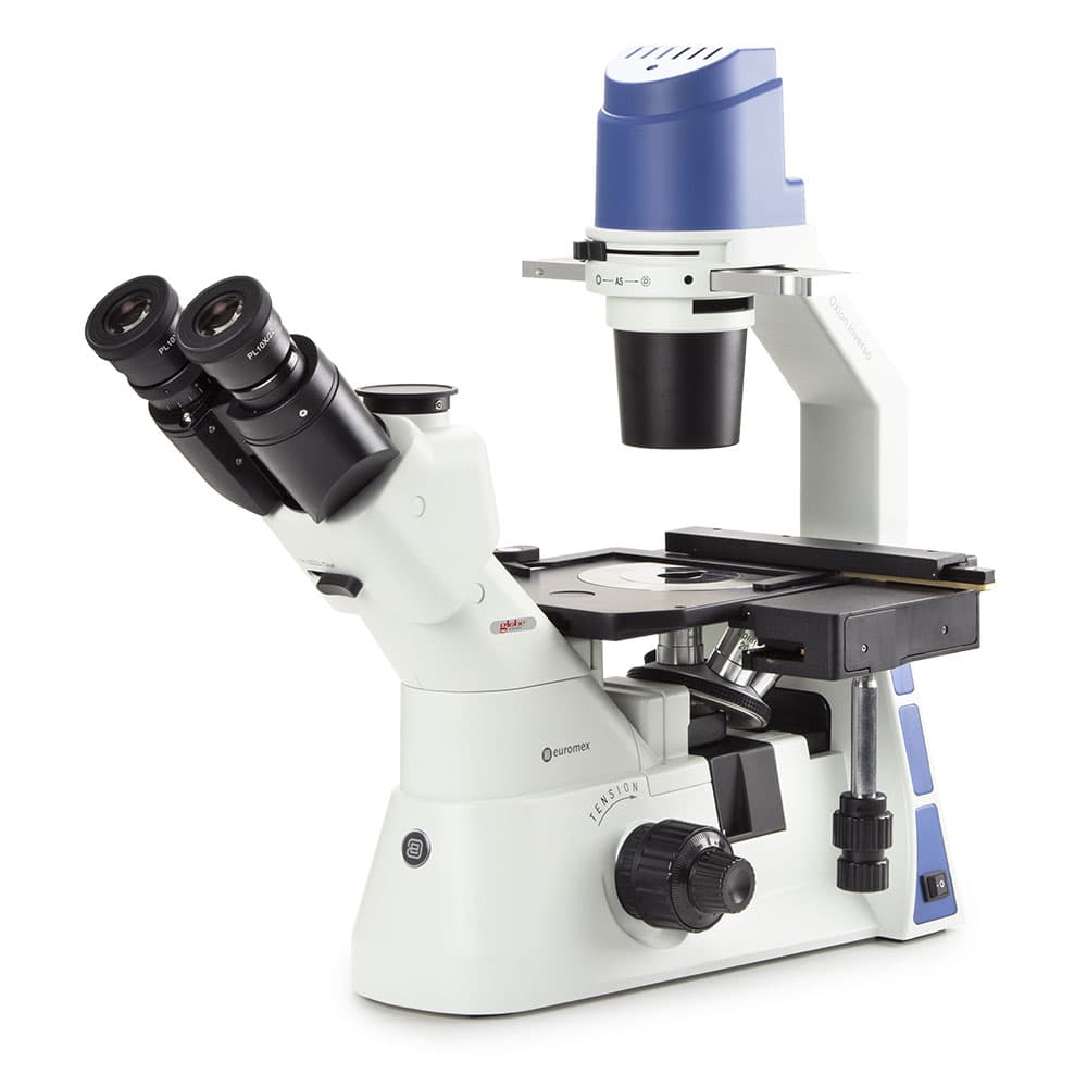
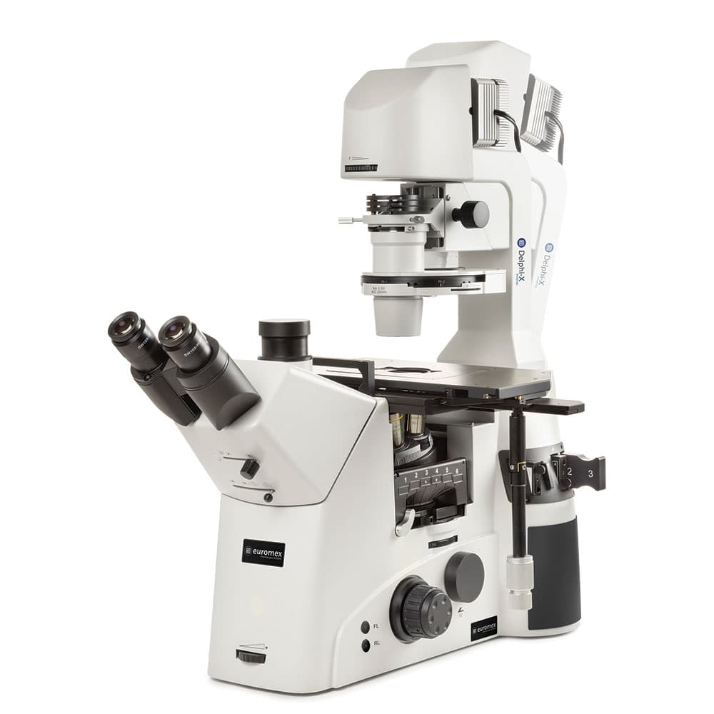
Showing 1 to 3 of 3 results
What Are Inverted Microscopes?
An inverted microscope is a compound light microscope in which the objective lens is positioned below the specimen stage, pointing upward, while the illumination system (condenser and light source) is above the stage, pointing downward. This inverted configuration allows cells growing on the inner floor of a culture dish, flask, or well plate to be observed directly through the clear plastic or glass base without removing the vessel lid and without disturbing the sterile culture. By contrast, upright microscopes require samples to be mounted on glass slides and covered with a coverslip for observation. Inverted microscopes are the universal tool for cell biology research; any workflow involving adherent mammalian cells, suspension cultures, organoids, embryos, or zebrafish in standard tissue culture vessels requires an inverted microscope for routine monitoring. Key contrast methods for live-cell observation are phase contrast (for unstained cells, the standard for assessing cell health, morphology, and confluency) and differential interference contrast (DIC, for optical sectioning). Fluorescence epi-illumination is added via a fluorescence port and filter cube turret for imaging GFP-labeled proteins, immunofluorescence-stained cells, or nuclear dyes such as DAPI. An inverted microscope is required for any workflow where cells are observed in their growth vessel (culture dishes, T-flasks, well plates, or spinner flasks). Phase contrast is non-negotiable for routine cell health monitoring; add fluorescence for reporter protein or immunofluorescence imaging.
What You Will Find:
- Inverted Trinocular Microscopes for imaging cells and cultures in dishes, flasks, and microplates.
- Mechanical Stages for precise and smooth sample positioning.
- Phase Contrast Optics for enhanced viewing of transparent, unstained cells.
- Advanced Imaging Systems with fluorescence-ready and DIC-prepared configurations.
- Camera-Compatible Trinocular Heads for digital imaging and documentation.
How to Choose an Inverted Microscope
Routine cell culture vs. research imaging
Routine cell culture microscopes prioritize phase contrast imaging, compact footprint, ease of use, and affordability; they are workhorses placed in biosafety cabinet rooms for daily culture monitoring. Research inverted microscopes add motorized stage and focus control, multi-channel LED fluorescence, automated XY scanning for multiwell plate imaging, programmable time-lapse, and software-controlled imaging modules for confocal, TIRF, and FRET; suited to imaging-centric research labs.
Phase contrast system
Phase contrast requires a matched pair of a phase objective and annular phase ring in the condenser. Low-contrast (PhI or Ph1) phase rings pair with low-power objectives (4x, 10x) for overview; medium-contrast (PhII or Ph2) rings pair with 20x and 40x objectives for standard cell morphology; high-contrast (PhIII or Ph3) rings pair with 60x or 100x oil-immersion objectives for high-magnification phase work. Confirm that the phase ring set installed in the condenser matches the objective phase designation for each magnification in use; mismatched pairs produce halos around cells rather than flat-field phase contrast.
Fluorescence: LED vs. mercury arc lamp
LED fluorescence illumination (available as an upgrade or built-in on most current inverted microscopes) provides 50,000+ hour lamp lifetime, instant-on/off switching (eliminating the warm-up and cool-down delays of mercury lamps), channel switching in less than 100 milliseconds for rapid multi-color imaging, and no mercury disposal requirement. Mercury arc lamps (HBO 50 or HBO 100) provide higher peak UV output for DAPI and Hoechst imaging, where LED UV intensity may be limiting, but require lamp replacement every 200 to 300 hours and strict mercury waste handling. For new purchases, LED fluorescence systems are the standard recommendation.
Objective working distance and culture vessel compatibility
Inverted microscope objective working distance (the distance from the front lens to the focused specimen) must exceed the thickness of the culture vessel base. Standard tissue culture polystyrene (PS) has a base thickness of 1 to 1.2 mm; glass-bottom dishes have 0.17 mm (No. 1.5 coverglass). Objectives specified for plastic culture vessels (abbreviated ELWD or LLWD) correct for the optical aberrations introduced by thick plastic and provide adequate working distance. Standard oil-immersion objectives are designed for No. 1.5 coverglass (0.17 mm) and cannot image through standard tissue culture plastic at high quality without significant spherical aberration.
Stage and sample holder
Confirm that the inverted microscope's stage accommodates your culture vessels: standard 35 mm and 60 mm culture dishes, T-25 and T-75 flasks, 6-well, 12-well, 24-well, 96-well, and 384-well plates. Universal stage holders with adjustable guides are available for most platforms; dedicated plate holders for 96-well and 384-well plates are used for high-content imaging applications. Motorized XY stages with encoder feedback are required for automated multiwell plate scanning and time-lapse experiments.
Specifications Context
For time-lapse live-cell imaging, focus drift over extended runs (hours to days) is the principal challenge; motorized focus with hardware autofocus maintains focus independent of thermal drift. Camera sensor size and pixel size determine the field of view at each magnification; a larger sensor captures a wider field per image, reducing the number of positions needed to tile a full well. For drug screening and plate-based assays, confirm that the software can drive the XY stage through a defined grid pattern and export images in a format compatible with your image analysis pipeline.
Bring your cellular research into focus—explore our range of inverted microscopes and contact the MBP team for a quote today.
FAQ