Microscope Accessories for Imaging and Stage Control
Microscope accessories expand the functionality of laboratory microscopes with components such as digital cameras for image capture, photo port adapters and C-mount adapters for camera integration, replacement camera tubes, and microscope illuminators for improved specimen lighting. Accessory compatibility is microscope-specific; always confirm the microscope model and manufacturer before ordering. Accessory compatibility is instrument-specific; confirm the microscope model and manufacturer before ordering any accessory.
MBP provides direct procurement support for microscope accessories for US and Canadian research laboratories. Request a quote for microscope accessories, including imaging systems, stage components, objectives, and fluorescence imaging upgrades by contacting customerservice@mbpinc.net.
Microscope Accessories
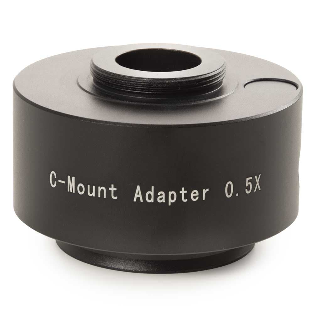
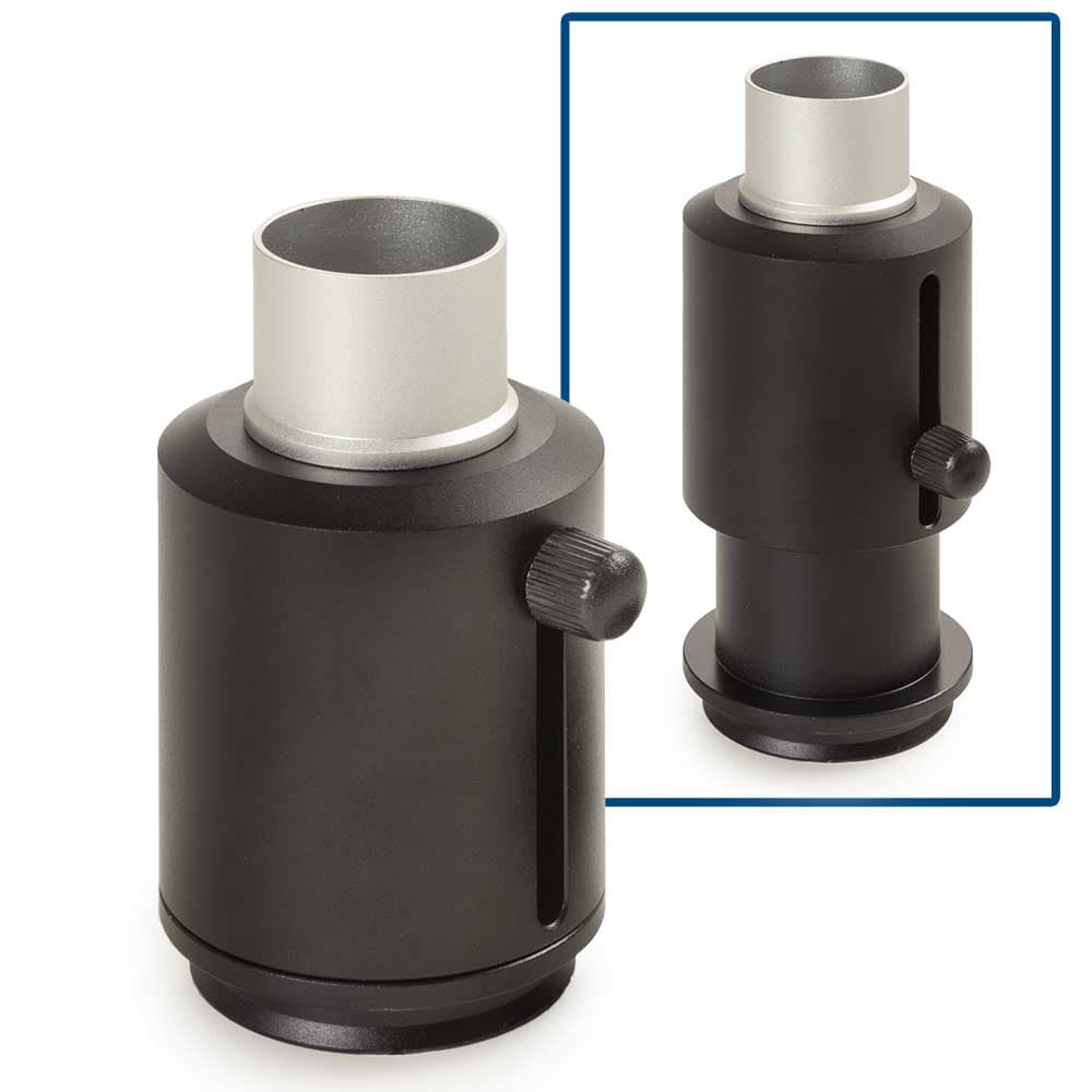
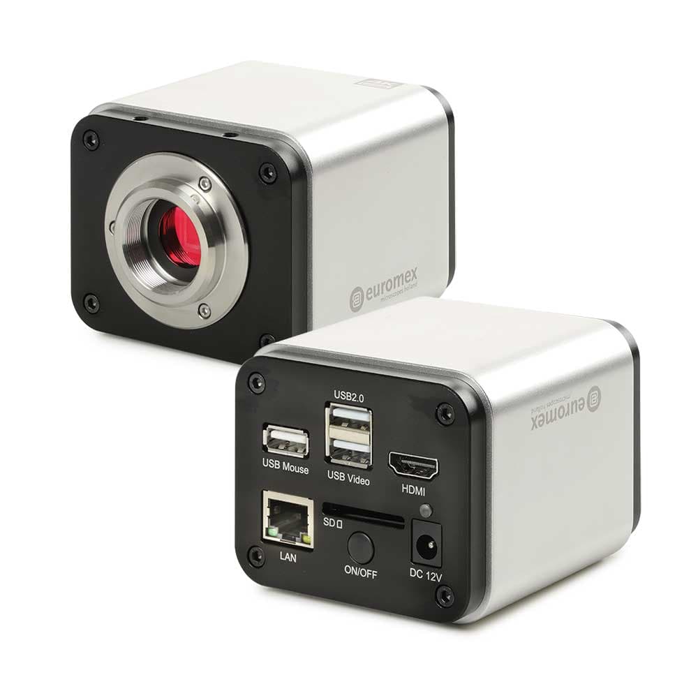
Showing 1 to 7 of 7 results
What Are Microscope Accessories?
Microscope accessories are supplementary components that configure, upgrade, or expand the function of a base microscope beyond its standard out-of-the-box specification. They fall into several categories: imaging accessories (cameras, camera adapters, camera relay lenses) for digital image capture and video documentation; optical accessories (additional or replacement objective lenses, eyepieces, auxiliary objectives for stereo microscopes) that change magnification or improve optical quality; fluorescence accessories (filter cube sets, LED fluorescence modules, mercury lamp adapters, UV/visible filter wheels) for adding or expanding fluorescence capability; stage accessories (multiwell plate holders, slide holders, cross-roller stages, motorized XY stage inserts, focus drives) that expand sample handling; illumination accessories (fiber optic illuminators, ring lights, polarizers, dark-field condensers) for alternative contrast methods; and maintenance and consumable accessories (immersion oil, lens paper, slide cleaning kits, replacement illuminator bulbs). Because accessory designs are instrument-specific (not all cameras, objectives, or stage inserts are universally compatible), identify the microscope model and manufacturer before ordering any accessory. Always identify the exact microscope model and manufacturer before ordering any accessory; thread standards, bayonet mounts, tube lengths, and connector formats vary significantly between brands and generations.
What You Will Find:
-
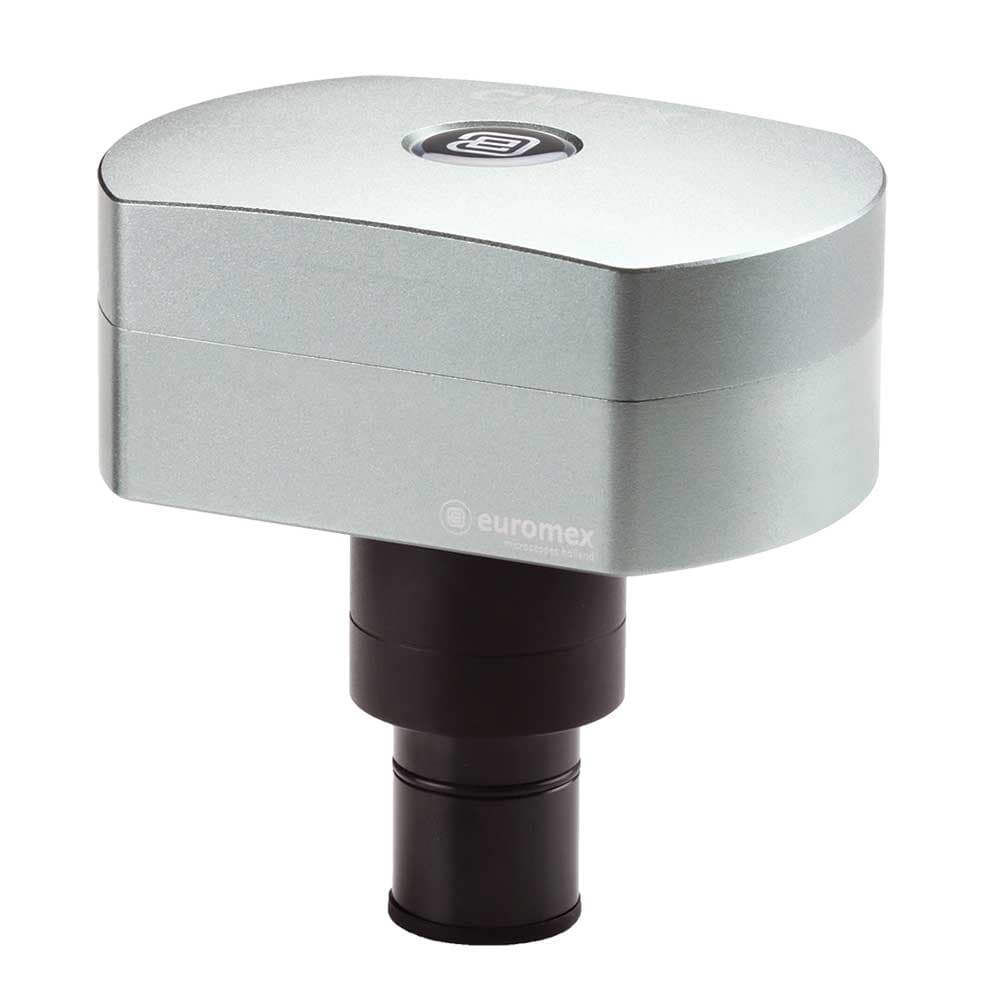
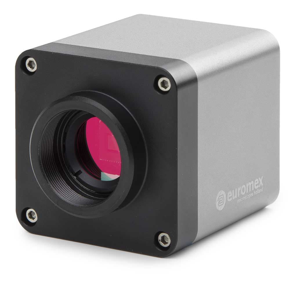
Microscope Cameras for high-resolution image and video capture, including HD and 4K imaging options.
-
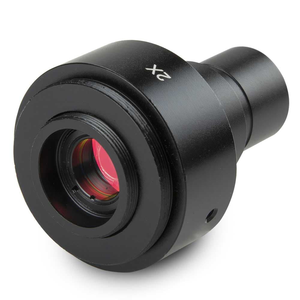
Photo Port Adapters and Camera Tubes for connecting digital cameras to compatible microscopes.
-
Microscope Adapters designed to support a wide range of imaging and mounting configurations.
-
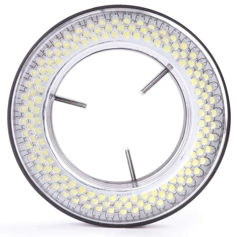
LED Illuminators that enhance specimen visibility and image quality across microscopy applications.
-
Digital Imaging Accessories that enable documentation, image analysis, teaching, and research workflows.
How to Choose Microscope Accessories
Digital cameras and adapters
Microscope cameras attach to the trinocular port (or monocular camera port on some models) via a C-mount camera adapter with an integrated relay lens. The relay lens magnification (typically 0.35x, 0.5x, or 1x) determines the effective field of view on the camera sensor. Choose a CMOS camera with 5 to 20 megapixels for brightfield documentation and routine fluorescence; scientific CMOS (sCMOS) or cooled CCD cameras for low-light fluorescence quantification. Confirm the camera software is compatible with the microscope's control software (if motorized).
Objective lenses
Replacement or additional objectives must match the microscope's objective thread standard and tube length (160 mm finite or infinity-corrected). Never mix finite-optics and infinity-corrected objectives on the same nosepiece; the image-forming properties are incompatible. Plan-apochromat objectives in the same brand family as the microscope are the safest choice for optical performance and parfocality with the existing objective set.
Fluorescence filter cube sets
Fluorescence filter cubes contain an excitation bandpass filter, a dichroic mirror, and an emission bandpass filter, which are the three components that define which fluorescent dye or protein is imaged. Common filter sets: DAPI (Ex 360/40, Di 400LP, Em 460/50); FITC/GFP (Ex 470/40, Di 495LP, Em 525/50); TRITC/mCherry (Ex 550/25, Di 565LP, Em 605/70); Cy5 (Ex 620/60, Di 660LP, Em 700/75). Confirm the cube block type (Olympus U-MF2, Nikon B-2A, Zeiss 38HE, or other) matches the filter cube turret of the specific microscope model.
Stage inserts and sample holders
Stage inserts expand sample compatibility: multiwell plate holders (96-well, 384-well, 6-well) for high-content imaging; glass-bottom dish holders for live-cell imaging with oil-immersion objectives; slide holder adapters for larger or smaller slide formats; and universal stage inserts for non-standard containers. Confirm the stage insert dimensions match the microscope's mechanical stage cutout and that the height profile is compatible with the condenser working distance for transmitted illumination.
LED fluorescence modules
LED fluorescence modules replace or augment mercury arc lamps in the fluorescence port of existing microscopes, providing individual LED channels for DAPI, GFP, TRITC, and Cy5 excitation. LED upgrades are available as bolt-on add-ons for many research-grade inverted and upright microscopes and significantly extend the fluorescence illumination service life from 200 to 300 hours (mercury) to 50,000+ hours (LED). Confirm the LED module's mounting standard and control interface (USB, TTL, or software-triggered) is compatible with the microscope.
Specifications Context
Immersion oil for oil-immersion microscopy must match the refractive index for which the objective is designed; standard immersion oil has a refractive index of 1.515 at 23 degrees C (per ASTM and DIN standards). Using oils with different refractive indices introduces spherical aberration. Viscosity matters for the application: low-viscosity oils (Type A, 150 mPas) spread easily and are used for fast scanning; high-viscosity oils (Type B, 1250 mPas) resist creep at elevated stage temperatures. Clean all residual immersion oil from objectives immediately after each use with lens tissue dampened with appropriate lens cleaning solution; dried immersion oil is difficult to remove and can permanently damage the objective coating.
To discuss product availability or get selection guidance, contact the MBP team.
FAQ