Gel Stains for Nucleic Acid and Protein Visualization
Gel Stains brings together the reagents used to detect nucleic acid and protein bands in a gel electrophoresis matrix: pre-staining and casting reagents incorporated into the gel before the run, post-staining reagents applied by soaking after electrophoresis, and loading dyes mixed with samples before loading. These are the three practical staining approaches every gel laboratory routinely chooses between based on convenience, safety, sensitivity, and fragment sizing accuracy. Academic and core laboratories selecting a staining strategy can benefit from guidance when comparing visualization methods and choosing reagents appropriate for their workflow.
Explore available gel stains or request a quotation by contacting customerservice@mbpinc.net. Our team can help identify the appropriate staining format and detection method for your nucleic acid or protein electrophoresis applications.
Gel Stains


Showing 1 to 6 of 6 results
What are gel stains?
Gel stains are the reagents used to make nucleic acid or protein bands visible in an electrophoresis gel or on a membrane: pre-staining reagents incorporated into the agarose before casting, post-staining reagents applied by soaking the gel after electrophoresis, and loading dyes mixed with samples before well loading. The most widely used nucleic acid stains are fluorescent DNA-intercalating dyes detected under UV or blue light, while the most common protein gel stains are Coomassie Brilliant Blue and silver stain for general protein detection.
What you will find:
-
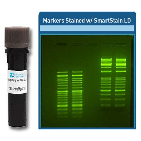
Loading Dye: for DNA visualization and sample preparation for electrophoresis, and includes Azura SmartStain Loading Dye, SmartGlow™ Loading Dye with Safe Green Stain.
-
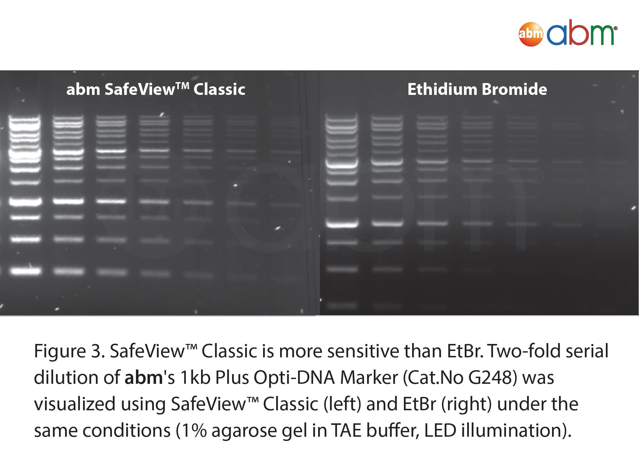
Pre-Stain/Casting: for nucleic acid detection and gel casting workflows. Examples include Azura SmartStain Pre-Stain, BlinkBlue SDS-PAGE Staining Buffer, and SafeView Classic.
-
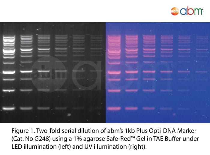
Post-Stain: for sensitive nucleic acid analysis and DNA visualization in electrophoresis. It includes a Safe-Red Gel stain.
How to choose gel staining approach
Choose pre-staining (casting) for convenience and real-time monitoring
Adding a fluorescent intercalating dye like ethidium bromide, GelRed, or SYBR Safe to the agarose before casting lets you visualize DNA bands as they migrate during electrophoresis and immediately after the run without a separate staining step, at the cost of some potential effect on DNA mobility from dyes incorporated into the gel matrix.
Choose post-staining when migration accuracy is most important
Post-staining by soaking the completed gel in stain solution avoids any effect on DNA migration during electrophoresis, since the stain is not present in the gel during the run, and consistently gives cleaner band backgrounds than some pre-staining methods. GelRed and SYBR Green are frequently recommended for post-staining for their superior sensitivity.
Use loading dye in every lane regardless of staining method
Loading dye is added to every DNA or RNA sample and to most ready-to-load ladders before loading to provide sample density for sinking into wells and a visible dye front for monitoring migration, independent of whichever nucleic acid stain is used to detect bands after or during electrophoresis.
Choose your nucleic acid stain based on UV versus blue-light visualization
Ethidium bromide and GelRed are detected with a standard UV (300–365 nm) transilluminator and EtBr filter; GelGreen and SYBR Safe can be detected under either UV or blue light, making them compatible with safer blue-light excitation systems for gel excision and downstream use of DNA.
For protein gels, select Coomassie stain for routine detection or silver stain for high sensitivity
Coomassie Brilliant Blue staining is the most common post-staining method for polyacrylamide protein gels, while silver staining provides substantially higher sensitivity but is more technically demanding and more easily compromised by background variation. Fluorescent protein stains are also available for compatibility with multi-channel fluorescent imaging systems.
Specifications context
Ethidium bromide, an intercalating dye that fluoresces strongly when bound to double-stranded DNA under UV light, is known to be mutagenic and requires specific waste disposal procedures, which has driven adoption of safer alternatives like GelRed and SYBR Safe that have similar UV-based excitation but significantly better safety profiles. As of 2026, GelRed and SYBR Safe are the most commonly adopted EtBr alternatives in labs transitioning away from ethidium bromide, with GelRed generally showing superior sensitivity, particularly in post-staining applications.
Contact the expert team at MBP and get premium-quality gel stains for your lab.
FAQ