Electrophoresis and Visualization Equipment for DNA, RNA, and Protein
Electrophoresis and gel visualization equipment are the core instruments for separating and detecting nucleic acids and proteins in molecular biology labs - encompassing horizontal gel systems for DNA and RNA separation, vertical SDS-PAGE systems for proteins, UV and blue-light transilluminators for fluorescent band visualization, and gel documentation imaging systems for digital recording and quantitative band analysis.
MBP supplies electrophoresis and visualization instruments from Biologix and Azura Genomics, compatible with standard agarose and polyacrylamide gel workflows, with US order processing in Houston, Texas, and shipping across North America and internationally. Request a quote by contacting customerservice@mbpinc.net.
Electrophoresis & Visualization



Showing 1 to 9 of 9 results
What Are Electrophoresis and Visualization Systems?
Electrophoresis and visualization systems are the paired instruments that separate biomolecules by size in a gel matrix under an electric field, then detect and record the separated bands using fluorescent dyes and light sources. Horizontal submarine agarose electrophoresis separates DNA and RNA fragments of 100 bp to 20 kb based on size; polyacrylamide (PAGE) separates smaller fragments or proteins with higher resolution. Visualization requires a transilluminator matched to the dye's excitation wavelength (UV at 302-365 nm for ethidium bromide; blue LED at 470 nm for SYBR Safe, GelGreen, SafeView) and, optionally, a gel documentation system to capture digital images. Choose electrophoresis and visualization equipment as a paired system: the gel tray, buffer system, power supply, stain, and imaging device must be compatible to ensure reproducible, publication-quality results.
What You Will Find:
-
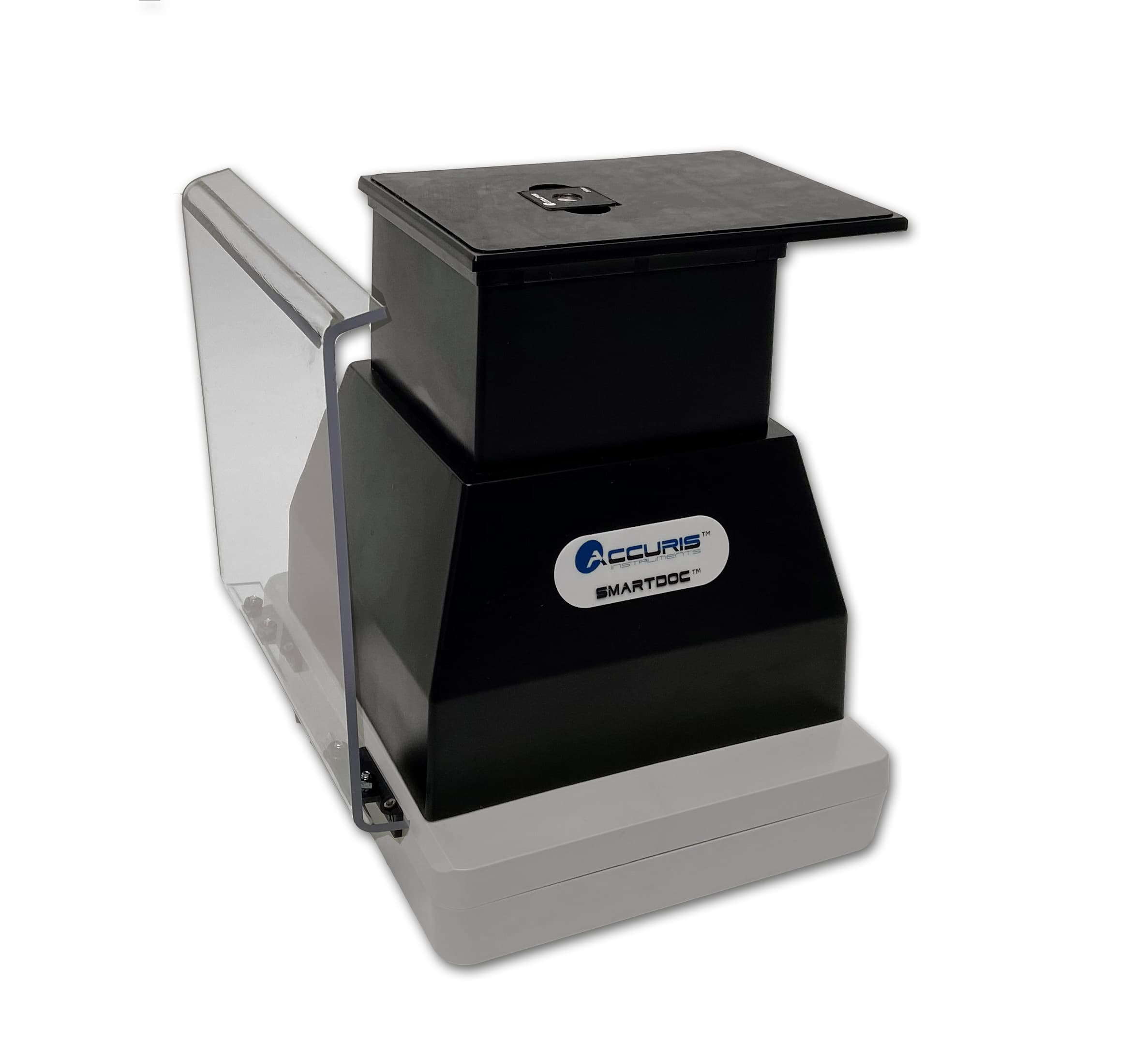
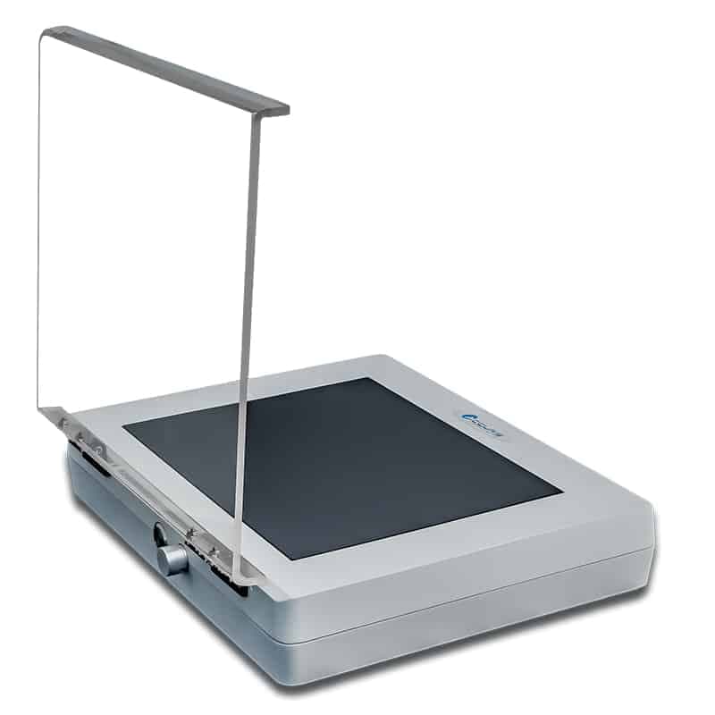
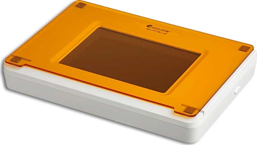
UV Transilluminators – High-intensity, uniform light sources designed for the clear and safe excitation of fluorescently stained gels.
-
Imaging and Gel Visualization – Advanced documentation systems that provide high-resolution captures and precise analysis of molecular bands.
-
Electrophoresis Systems – Robust horizontal and vertical tanks engineered for consistent current flow and superior separation of nucleic acids and proteins.
How to Choose Electrophoresis and Visualization Equipment
Nucleic acid vs. protein electrophoresis
Horizontal agarose gel electrophoresis (0.5-3% agarose in TAE or TBE buffer) resolves DNA and RNA fragments from ~100 bp to 20 kb and is the standard for PCR product verification, restriction digest analysis, and RNA integrity assessment. Vertical polyacrylamide gel electrophoresis (SDS-PAGE, 8-15% acrylamide) separates proteins from 10 to 200 kDa with higher resolution than agarose and is required for western blotting. For oligonucleotide or small RNA analysis below 100 bp, a denaturing polyacrylamide gel (TBE-urea) provides the necessary resolution.
Gel tray size and well count
Mini trays (7 x 8 cm, 8-16 wells) suit quick PCR checks with fewer than 16 samples. Standard trays (10 x 14 cm, 20-30 wells) handle routine molecular biology workflows. Wide-format trays (20 x 20 cm, 96+ wells) support high-throughput colony screens. Multi-comb systems allow variable lane configurations on the same tray. Well volume capacity (10-50 microliters per well) should match your standard sample loading volume.
Stain and transilluminator compatibility
Ethidium bromide (EtBr, excitation 302 nm, emission 590 nm) requires UV transilluminators at 302-365 nm and standard orange/amber photographic filters. EtBr is mutagenic and requires dedicated disposal. Safer alternatives, including SafeView (Safe-Green: 490/525 nm; Safe-Red: 540/630 nm), SYBR Safe (excitation ~300 nm and 497 nm, emission 530 nm), and GelGreen, are visualized with blue-LED transilluminators at 470 nm and are compatible with standard disposal. SafeView stains are available directly from MBP and have a sensitivity of 0.2-0.8 ng per band.
Gel documentation system requirements
For basic agarose gel recording, a 5-8 megapixel camera gel doc system provides sufficient resolution for PCR band documentation. For quantitative western blot densitometry or low-abundance band detection, choose a 12-20 megapixel system with 16-bit dynamic range, which resolves bands at 0.1 ng or below. Software integration with analysis packages (ImageJ-compatible export, TIFF output) is essential for publication-grade figures.
Power supply integration
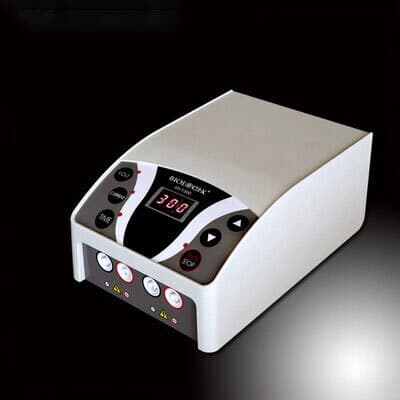
Electrophoresis power supplies should be purchased alongside the gel system. Standard agarose gel electrophoresis operates at 80-150 V constant voltage; mini gels typically run at 80-100 V for 30-45 minutes. Ensure the power supply can drive multiple gel units simultaneously if throughput is a priority, with a combined current capacity of 400-500 mA.
Specifications Context
Agarose concentration determines the size range resolved: 0.5-0.8% agarose resolves 1-20 kb DNA fragments; 1-1.5% resolves 200 bp-10 kb; 2-3% resolves 50-500 bp. TAE (Tris-acetate-EDTA) buffer is standard for agarose gel electrophoresis and cloning applications; TBE (Tris-borate-EDTA) gives sharper bands for smaller fragments below 1 kb. UV exposure of DNA in agarose gels for more than 30 seconds at 302 nm can reduce cloning efficiency significantly; blue-light transilluminators at 470 nm cause minimal DNA damage, preserving viability for downstream gel extraction and cloning. Blue-LED transilluminators with LED panels rated for 50,000+ hours have largely replaced fluorescent UV tubes in new lab installations, reducing running costs and eliminating the need for UV tube replacement every 2-3 years.
Don’t let your bands go unnoticed. Browse our electrophoresis range and hit up the MBP team for a custom quote today.
FAQ