RNA Tracking Dyes for Live-Cell RNA Visualization and Localization Studies
RNA tracking dyes are cell-permeant, fluorogenic small molecules that selectively bind RNA over DNA, lighting up only when bound to give a clean signal for visualizing RNA distribution in living cells without genetic labeling or fixation. They are used for tracking RNA granule transport in neurons, visualizing RNA cargo in exosomes, and resolving nucleolar structure, since the dye is virtually nonfluorescent until it binds its target. Academic and core laboratories studying RNA localization, trafficking, and dynamics in live cells can benefit from specialist guidance when selecting dyes based on fluorescence properties, live-cell compatibility, and downstream imaging requirements.
Explore available RNA tracking dyes or request a quotation by contacting customerservice@mbpinc.net. Our team can help identify the appropriate RNA-selective dye and imaging workflow for your live-cell RNA visualization applications.
RNA Tracking dyes
Showing 1 to 4 of 4 results
What are RNA tracking dyes?
RNA tracking dyes are cell-permeant, fluorogenic nucleic acid stains that selectively bind RNA over DNA, remaining virtually nonfluorescent in the unbound state and lighting up brightly once bound to RNA, which gives a clean, low-background signal for live-cell imaging. The best-characterized commercial example binds RNA with absorption and emission maxima around 490/530 nm and only weak signal when bound to DNA, validated by RNase and DNase removal experiments showing that RNase treatment eliminates nucleolar and cytoplasmic labeling while DNase treatment does not.
What you will find:
-
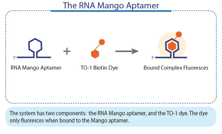
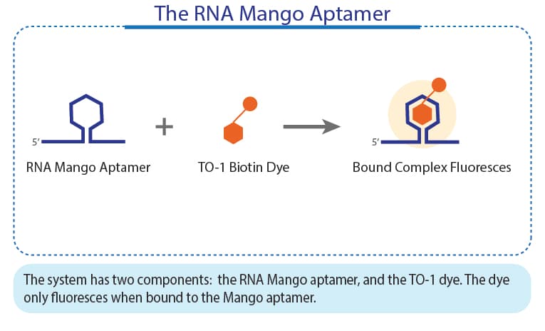
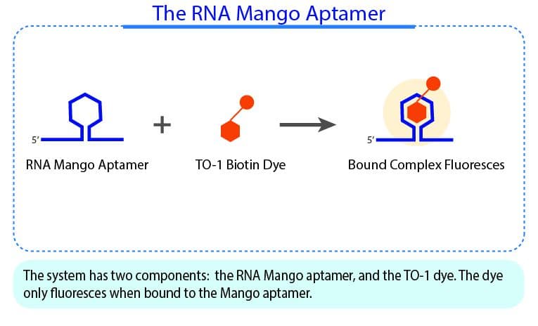
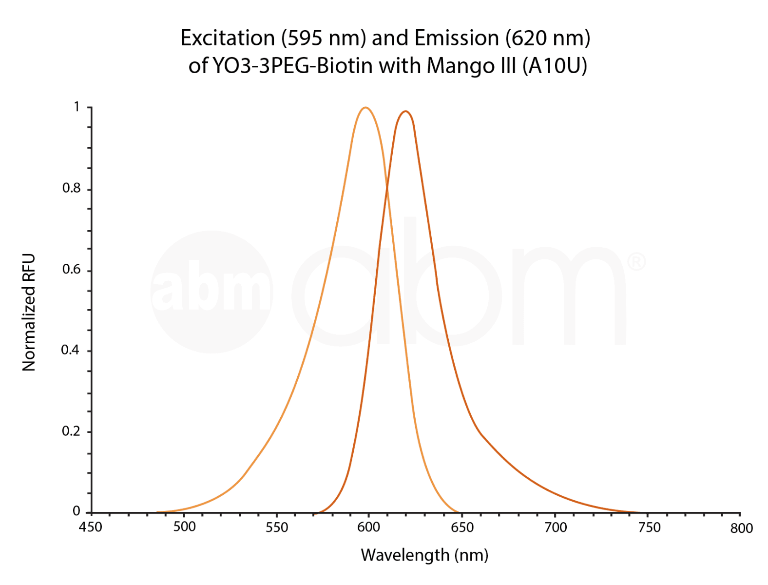
Biotinylated Fluorophores: High-affinity markers such as TO1, TO3, and YO3-3PEG-Biotin, engineered for robust labeling and permanent capture of RNA molecules in various structural and functional assays.
-
Desthiobiotin Modifications: Specialized tools like the TO1-3PEG-Desthiobiotin Fluorophore (G7956) that allow for reversible binding and gentle elution, making them ideal for enrichment workflows where maintaining the native state of the RNA is critical.
How to choose RNA tracking dyes
Confirm the dye is genuinely RNA-selective rather than a general nucleic acid stain
A true RNA-selective dye shows bright fluorescence when bound to RNA but only weak signal when bound to DNA, a distinction worth confirming against published spectral and RNase/DNase validation data before committing to a specific dye for an RNA-specific application.
Match the dye to your specific RNA visualization goal
Different dyes in a related family vary in their subcellular binding behavior, with some binding cytoplasmic RNA specifically, useful for tracking RNA granule transport in living neurons, while others are optimized for general nucleolar and cytoplasmic RNA staining.
Plan your fixation method around the dye's stated compatibility
Methanol fixation after staining generally preserves an RNA tracking dye's staining pattern with minimal loss, while formaldehyde fixation can alter the staining pattern, a distinction worth confirming before choosing a fixation protocol for downstream analysis.
Consider a dye sampler kit when working with an unfamiliar cell or tissue type
Dye staining behavior can vary across different cell and tissue types, so a sampler kit containing several related dyes lets you empirically determine which performs best for your specific sample before committing to a single dye for a full study.
Confirm buffer compatibility for the staining solution
RNA tracking dyes are generally compatible with standard physiological buffers in the pH 7.0–8.0 range, including PBS, for preparing the staining solution used in live-cell experiments.
Specifications context
RNA tracking dyes have been used to stain RNA cargo in exosomes in both live-cell samples and isolated exosome preparations, and to differentiate apoptotic from non-apoptotic cells when paired with other viability indicators, reflecting the breadth of live-cell RNA applications beyond simple visualization. As of 2026, newer experimental small-molecule RNA dyes reporting higher fluorogenic enhancement and improved photostability have been described in the academic literature as potential alternatives to the long-standing commercial standard, though these remain research-stage rather than established catalog items.
Visualize your transcripts with precision—contact the MBP team today for a quote on our professional RNA tracking solutions.
FAQ